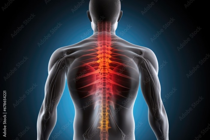
Back pain, a common ailment affecting millions, presents in diverse forms. Understanding the root cause is crucial for effective treatment. This exploration delves into three primary types: muscle strain, disc herniation, and spinal stenosis, examining their distinct characteristics, diagnostic methods, and differentiating factors. We’ll navigate the complexities of physical examinations, imaging techniques, and neurological assessments to provide a clearer picture of how these conditions are identified and managed.
By differentiating between these three types, we aim to highlight the importance of accurate diagnosis for appropriate treatment and pain management. This detailed analysis will empower individuals and healthcare professionals alike to better understand and address this prevalent health concern. We will explore the nuances of each condition, comparing symptoms, risk factors, and the most effective diagnostic approaches.
Understanding the Three Main Types of Back Pain
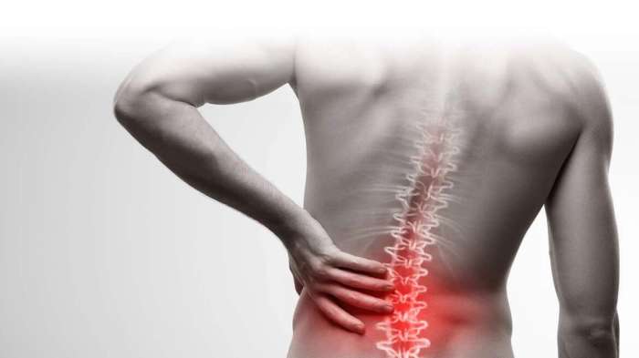
Back pain is a common ailment affecting people of all ages and activity levels. Understanding the different types of back pain is crucial for effective diagnosis and treatment. This section will detail three primary types: muscle strain, disc herniation, and spinal stenosis, focusing on the anatomical structures involved, pain characteristics, and typical onset and progression.
Anatomical Structures Involved in Back Pain
The spine is a complex structure composed of bones (vertebrae), discs, ligaments, muscles, and nerves. Each type of back pain affects specific structures within this intricate system, leading to unique symptoms and treatment approaches. The following table summarizes the affected structures, common symptoms, and potential causes for each of the three main types of back pain.
| Pain Type | Affected Structure | Common Symptoms | Potential Causes |
|---|---|---|---|
| Muscle Strain | Muscles, ligaments | Localized pain, muscle spasms, stiffness, tenderness to the touch. Pain is often aggravated by movement and relieved by rest. | Overexertion, poor posture, sudden movements, inadequate warm-up before exercise. |
| Disc Herniation | Intervertebral discs, nerves | Sharp, shooting pain that may radiate down the leg (sciatica), numbness, tingling, weakness in the leg or foot. Pain can be severe and debilitating. | Degeneration of the intervertebral disc, injury, repetitive strain, aging. |
| Spinal Stenosis | Vertebral bones, ligaments, spinal canal | Pain, numbness, tingling, weakness in the legs and buttocks. Pain is often worse with standing or walking and improves with sitting or bending forward. | Degeneration of the spine, bone spurs, thickened ligaments, aging. |
Differences in Pain Location and Characteristics
Muscle strain typically presents as localized pain in the back muscles, often felt as a dull ache or stiffness. The pain is usually aggravated by movement and relieved by rest. In contrast, disc herniation often causes sharp, radiating pain, commonly known as sciatica, that travels down the leg along the sciatic nerve. Spinal stenosis presents with pain, numbness, and weakness in the legs and buttocks, often exacerbated by standing or walking and relieved by sitting or bending forward. The pain’s location and characteristics help differentiate these conditions.
Typical Onset and Progression of Back Pain
Muscle strains often have a sudden onset, frequently following a specific event like lifting a heavy object or engaging in strenuous activity. The pain typically improves within a few days to weeks with rest and conservative treatment. Disc herniation can have a sudden onset, often triggered by a specific incident, or it may develop gradually over time. The pain can be severe and persistent, potentially requiring more extensive treatment. Spinal stenosis usually develops gradually over many years as a result of age-related degenerative changes in the spine. The pain and symptoms typically worsen over time, potentially requiring ongoing management.
Diagnosing Each Type of Back Pain
Accurately diagnosing the source of back pain is crucial for effective treatment. This involves a comprehensive approach combining a detailed patient history, a thorough physical examination, and, in many cases, appropriate imaging and neurological testing. The specific diagnostic methods employed will vary depending on the suspected type of back pain – mechanical, radicular, or spinal stenosis.
Physical Examination for Back Pain
A systematic physical examination is the cornerstone of diagnosing back pain. This allows clinicians to pinpoint the location, nature, and potential cause of the pain, guiding further investigations.
- Observation: The patient’s posture, gait, and any visible signs of muscle atrophy or asymmetry are noted.
- Palpation: The spine is palpated to identify areas of tenderness, muscle spasm, or bony abnormalities. This includes assessing the paraspinal muscles, spinous processes, and sacroiliac joints.
- Range of Motion: Active and passive range of motion of the spine is assessed in flexion, extension, lateral bending, and rotation. Limitations or pain during specific movements can indicate the source of the problem.
- Neurological Examination: This involves testing muscle strength, reflexes, and sensation in the lower extremities to detect any nerve root compression or spinal cord involvement (discussed further below).
- Special Tests: Specific tests, such as the straight leg raise test (to assess sciatic nerve irritation) and various other orthopedic tests, may be performed to further localize the pain source.
Role of Imaging Techniques in Diagnosing Back Pain
Imaging techniques play a vital role in visualizing the structures of the spine and identifying underlying pathologies. The choice of imaging modality depends on the clinical suspicion and the information needed.
| Imaging Technique | Mechanical Back Pain | Radicular Back Pain | Spinal Stenosis |
|---|---|---|---|
| X-ray | Useful for identifying fractures, spondylolisthesis, and other bony abnormalities. Often the initial imaging study. | May show degenerative changes but is less sensitive for nerve root compression. | Can reveal narrowing of the spinal canal, but MRI is generally preferred for detailed visualization. |
| MRI | Can visualize soft tissues (intervertebral discs, ligaments, muscles) and identify disc herniations, although often not necessary for uncomplicated cases. | Excellent for visualizing nerve root compression and identifying the level and extent of herniated discs. | Provides the best visualization of the spinal canal and nerve roots, allowing for precise assessment of stenosis. |
| CT Scan | Less frequently used than MRI for mechanical back pain, but can be helpful in identifying bony abnormalities or fractures in more detail. | Can be used to visualize nerve root compression, but MRI offers superior soft tissue contrast. | CT myelography (CT scan with contrast injected into the spinal canal) may be used in specific cases to better visualize the spinal cord and nerve roots. |
Neurological Tests for Back Pain
Neurological tests help determine if nerve roots are involved in the back pain. This is crucial for diagnosing radicular pain.
A structured neurological examination report might include:
Patient: [Patient Name], [Date]
Examination: Lower extremity neurological examination performed.
Findings:
- Muscle Strength: Right lower extremity strength 5/5; left lower extremity strength 4/5 in the tibialis anterior muscle (suggestive of L4 nerve root involvement).
- Reflexes: Patellar reflexes 2+ bilaterally; Achilles reflexes 1+ on the left (hyporeflexia suggestive of nerve root involvement).
- Sensation: Decreased sensation to light touch and pinprick along the L4 dermatome on the left leg.
- Straight Leg Raise Test: Positive on the left at 45 degrees.
Impression: Findings suggest left L4 nerve root compression.
Differentiating Between the Three Types of Back Pain
Accurately diagnosing the source of back pain is crucial for effective treatment. While muscle strain, disc herniation, and spinal stenosis all manifest as back pain, their underlying mechanisms, symptoms, and prognoses differ significantly. Understanding these distinctions is key to guiding appropriate interventions.
Comparison of Symptoms
The following table highlights key differences in the presentation of muscle strain, disc herniation, and spinal stenosis. Note that symptom overlap can occur, making accurate diagnosis challenging.
| Feature | Muscle Strain | Disc Herniation | Spinal Stenosis |
|---|---|---|---|
| Pain Location | Localized to the affected muscle group; often in the lower back | Lower back pain, often radiating down one leg (sciatica) following a dermatomal pattern | Lower back pain, often radiating down both legs (bilateral); pain may worsen with prolonged standing or walking |
| Pain Character | Aching, cramping; may be sharp with sudden movement | Sharp, shooting, burning pain; can be intense | Dull, aching pain; may be accompanied by numbness, tingling, or weakness in the legs |
| Onset | Gradual onset, often related to overuse or strain | Sudden onset, often related to a specific event like lifting a heavy object | Gradual onset, often worsening over time |
| Physical Exam Findings | Muscle tenderness, spasm, decreased range of motion | Positive straight leg raise test, decreased reflexes, muscle weakness | Decreased range of motion, neurological deficits (e.g., weakness, sensory loss) in the legs, positive neurologic tests |
Scenarios of Mimicry and Differentiation
Muscle strains can sometimes mimic the early stages of disc herniation, presenting with similar lower back pain. However, the absence of radiating pain (sciatica) and neurological deficits in muscle strain helps differentiate it. Similarly, spinal stenosis can initially present with symptoms resembling a severe muscle strain. However, the progressive nature of spinal stenosis, its bilateral leg pain, and the presence of neurological symptoms will eventually distinguish it. Detailed physical examination, including neurological assessments and imaging studies (MRI or CT scans), are critical for accurate diagnosis in these ambiguous cases.
Risk Factors for Each Type of Back Pain
Understanding the risk factors for each type of back pain can aid in preventative measures and early diagnosis.
Several factors increase the likelihood of developing each type of back pain:
- Muscle Strain: Poor posture, lack of physical fitness, sudden strenuous activity, muscle imbalances, and previous injuries are significant risk factors. For example, a construction worker repeatedly lifting heavy objects without proper lifting techniques is at high risk for muscle strain.
- Disc Herniation: Age-related degeneration of the intervertebral discs, repetitive heavy lifting, poor posture, and genetic predisposition all increase the risk. A person with a family history of disc herniation and a job involving frequent bending and lifting is at increased risk.
- Spinal Stenosis: Aging is a primary risk factor, leading to the gradual narrowing of the spinal canal. Osteoarthritis, spondylolisthesis (a slippage of one vertebra over another), and certain genetic conditions also increase susceptibility. An elderly individual with a history of osteoarthritis and spondylolisthesis is at significantly higher risk.
Final Conclusion

Accurate diagnosis is paramount in effectively managing back pain. This comprehensive overview of muscle strain, disc herniation, and spinal stenosis has illuminated the key differences in their presentation, diagnostic methods, and risk factors. By understanding these distinctions, healthcare providers can tailor treatment plans to individual needs, leading to improved patient outcomes and a better quality of life. Remember, seeking professional medical advice is crucial for any persistent back pain.